In the medical field, the application of a beam in medical imaging plays a crucial role in diagnostics and treatment planning.
What Is a Beam in Medical Imaging?
A beam in medical imaging refers to a focused stream of energy that is utilized in various imaging modalities to create visual representations of the inside of the body.
These beams can manifest in several forms, including:
- X-rays
- Ultrasound waves
- Magnetic resonance imaging fields
- Gamma rays
Each type serves its unique purpose and applications in diagnostics. Understanding how these beams work allows healthcare professionals to leverage their capabilities effectively.
How Does Each Technology Use Beams?
X-ray Radiography
X-ray radiography is one of the most common applications of a beam in medical imaging.
- X-rays pass through the body and are absorbed at different rates by various tissues.
- Dense materials such as bones absorb more X-rays, resulting in darker images, while softer tissues appear lighter.
This allows radiologists to evaluate fractures, infections, or tumors. X-ray beams are not only quick to capture, but they also require minimal patient preparation, making them a go-to option in urgent care environments.
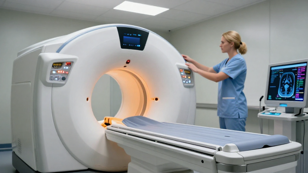
Computed Tomography (CT)
Computed Tomography (CT) scans elevate the concept of a beam in medical imaging to new heights.
- CT uses a rotating X-ray beam to obtain cross-sectional images of the body.
- This method allows for 3D reconstructions and offers more detailed imagery than standard X-rays.
Because CT scans provide comprehensive information, they are often indispensable in the diagnosis of complex medical conditions, including internal injuries and cancers.
Magnetic Resonance Imaging (MRI)
The application of a beam in medical imaging extends to Magnetic Resonance Imaging (MRI) as well.
- MRI uses a strong magnetic field combined with radiofrequency waves that generate detailed images of organs and tissues.
- Instead of using ionizing radiation, MRI leverages magnetic beams, making it safer for repeated examinations.
MRI is particularly valuable for visualizing soft tissues like the brain, spinal cord, and muscles, where precision is paramount.
Ultrasound Imaging
Ultrasound imaging is another area where a beam in medical imaging is effectively utilized.
- Here, high-frequency sound waves are emitted via a transducer, creating images based on the echoes that bounce back from tissues.
- This technique is primarily used for monitoring fetal development, as well as examining abdominal organs.
Ultrasound is favorable because it is non-invasive, has no known health risks, and can produce real-time images.
Positron Emission Tomography (PET)
Positron Emission Tomography (PET) involves the use of gamma-ray beams to visualize metabolic activity in tissues.
- Patients are administered a small amount of radioactive material that emits gamma rays.
- Special cameras pick up these rays, allowing for the detailed imagery of organ function.
PET scans are widely used in oncology to detect cancer and to evaluate treatment effectiveness through metabolic or physiological changes.
What Are the Advantages of Using Beams in Medical Imaging?
The use of beams in medical imaging comes with several key advantages:
-
Non-invasiveness: Most imaging techniques allow for internal evaluation without the need for surgery or incisions.
-
Detailed Visualization: Various beams provide high-resolution images, improving diagnostic accuracy.
-
Real-time Feedback: Techniques like ultrasound provide instant feedback, which is crucial in emergency situations.
-
Comprehensive Diagnostic Capabilities: Different types of beams serve various diagnostic needs, covering a wide range of conditions.
Are There Risks Associated With Beams in Medical Imaging?
While the benefits of using a beam in medical imaging are immense, it is essential to consider potential risks, particularly with specific modalities.
-
Radiation Exposure: X-rays and CT scans involve exposure to ionizing radiation, which can increase cancer risk if used excessively.
-
Allergic Reactions: In some cases, patients may have allergic reactions to contrast agents used in CT or MRI procedures.
-
Environmental Concerns: The production and disposal of radioactive materials used in modalities like PET raise environmental concerns.
Therefore, informed consent and careful consideration of the risks vs. benefits are paramount for safe medical imaging practices.
How Are Beams Being Improved in Medical Imaging Technologies?
Technological advancements continuously enhance the role of beams in medical imaging. Current trends focus on:
-
Increasing resolution: Developments in detector materials and imaging algorithms allow for clearer images.
-
Reducing exposure: Innovations aim to minimize radiation exposure during X-ray and CT imaging.
-
Integrating AI: Artificial Intelligence is being utilized for more accurate detection and diagnosis, thereby optimizing the analysis of imaging data.
-
Portable imaging devices: Researchers are working on compact, portable imaging machines that can deliver the benefits of traditional machines without being cumbersome.
Conclusion: The Future of Beams in Medical Imaging
The role of a beam in medical imaging is indispensable for modern diagnostics.
From X-rays to MRIs, various forms of beams provide insights into human health that were once impossible.
With ongoing advancements in technology, these imaging modalities are becoming more efficient, safer, and less invasive.
As we move forward, the integration of innovative approaches and technologies will revolutionize how medical professionals use beams in imaging, ultimately leading to better patient outcomes.
In sum, understanding the versatile applications of beams in medical imaging will not only improve diagnostics and treatments but also pave the way for groundbreaking advancements in the future.