In the world of medical and industrial imaging, beam imaging techniques play a crucial role in enabling detailed examinations and analyses. This article explores the various types of beam imaging techniques, their applications, and how they work.
What Are Beam Imaging Techniques?
Beam imaging techniques refer to methods that utilize beams of energy, such as X-rays, gamma rays, or electrons, to create images of internal structures. These techniques are foundational in both medical diagnostics and many industrial applications.
Why Are Beam Imaging Techniques Important?
- Non-Invasive: They allow for examination without surgical intervention.
- High Resolution: These techniques provide detailed images.
- Real-Time Analysis: Many techniques allow for the real-time assessment of structures.
- Versatile Applications: Used in various fields, including healthcare, engineering, and security.
Which Types of Beam Imaging Techniques Exist?
Several beam imaging techniques stand out in terms of their application and effectiveness.
What is X-ray Imaging?
X-ray imaging is one of the most common beam imaging techniques used in medical settings.
- What it Uses: X-ray beams to penetrate tissues.
- Applications: Detects fractures, infections, and tumors.
- Advantages:
- Quick and efficient.
- Provides immediate results.
- Available in most healthcare facilities.
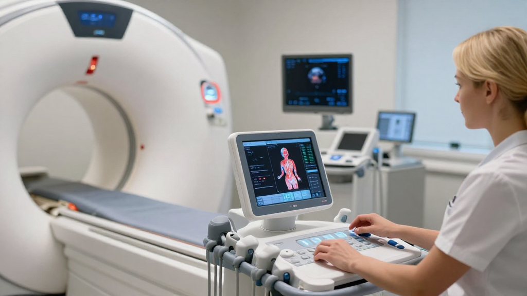
How Does Computed Tomography (CT) Work?
Computed Tomography (CT) is an advanced imaging technique that combines multiple X-ray images.
- What it Uses: A rotating beam of X-rays.
- Applications: Comprehensive imaging of abdominal organs, bones, and the brain.
- Advantages:
- Produces cross-sectional images.
- Provides precise anatomical information.
- Useful for diagnosing complex conditions.
What About Positron Emission Tomography (PET)?
Positron Emission Tomography (PET) employs a different approach to imaging.
- What it Uses: Gamma rays emitted from radioactive tracers.
- Applications: Often used in oncology to detect cancer and monitor treatment response.
- Advantages:
- Shows metabolic activity.
- Can identify diseases earlier than other imaging techniques.
What is Fluoroscopy?
Fluoroscopy is an imaging technique that allows real-time visualization of internal structures.
- What it Uses: Continuous X-ray beams.
- Applications: Often used for guiding surgical procedures or examining the gastrointestinal tract.
- Advantages:
- Live action images.
- Assists in dynamic assessments.
How is Ultrasound Related?
While typically not categorized with beam imaging techniques using ionizing radiation, ultrasound does utilize beams.
- What it Uses: High-frequency sound waves.
- Applications: Common in prenatal imaging and assessing soft tissues.
- Advantages:
- Safe and non-invasive.
- No exposure to radiation.
Can Magnetic Resonance Imaging (MRI) Use Beams?
Though magnetic resonance imaging does not primarily utilize beams in the same way X-rays do, it can be indirectly associated with beam imaging techniques.
- What it Uses: Magnetic fields and radio waves.
- Applications: Ideal for soft tissue imaging, including muscles and organs.
- Advantages:
- Highly detailed images.
- No ionizing radiation.
How Do Beam Imaging Techniques Work?
Understanding the operational principles behind beam imaging techniques enriches our appreciation of their capabilities.
What is the Basic Principle of Beam Interaction?
At the core, these imaging techniques rely on the interaction of beams with matter:
- Absorption: Different tissues absorb X-rays or gamma rays to varying degrees.
- Refraction: Some beams are bent in their passage through different media.
- Scattering: Beams can scatter, providing additional information about the internal structure.
What Equipment is Used for Beam Imaging?
Each technique relies on specialized equipment designed to deliver and capture beam interactions:
- X-ray machines: Produce and detect X-rays.
- CT scanners: Combines X-ray data using computerized algorithms.
- PET scanners: Detect gamma rays emitted from radiotracers.
- Fluoroscopy machines: Provide real-time imaging using X-ray technology.
Are There Risks Involved with Beam Imaging Techniques?
Safety is essential in any medical procedure, including beam imaging techniques.
What Are the Potential Risks?
- Radiation Exposure: X-rays and gamma rays are forms of ionizing radiation that can pose risks.
- Allergic Reactions: Some procedures may require contrast agents, which may cause allergies in some individuals.
- Misinterpretation: Images may sometimes be misinterpreted, leading to incorrect diagnoses.
How Can Risks Be Minimized?
- Proper Training: Ensure that technicians and radiologists are thoroughly trained.
- Patient History: Always evaluate patient history and potential allergies before administering imaging.
- Latest Equipment: Use updated technology that provides the lowest possible radiation doses.
Conclusion
In summary, beam imaging techniques are integral to various fields, providing vital information for diagnosis and analysis.
From X-ray imaging to PET scans, each method offers unique benefits and applications.
By understanding the intricacies of how these techniques work, we can appreciate the remarkable ability they give us to peek inside the human body and other complex structures.
Further Considerations
As technology advances, the future of beam imaging techniques looks promising. Emerging technologies such as photoacoustic imaging and hybrid imaging systems could redefine how we approach medical diagnostics and industrial safety.
Navigating the world of beam imaging not only enhances our knowledge of technology but also empowers us to make informed decisions regarding healthcare and engineering applications.